Double Floor Of Sella Turcica
Double Sellar Floor Sign A Clue Of Pituitary Tumor Springerlink
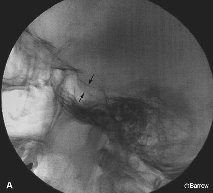
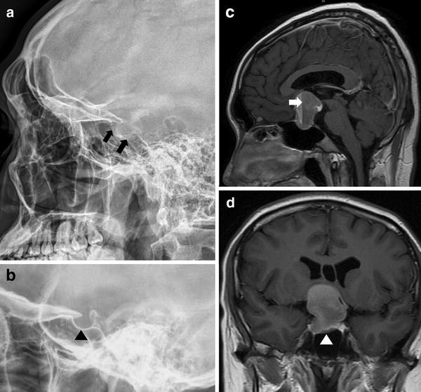
Double Sellar Floor Radiographic Sign For A Pituitary Adenoma Barrow

Specialised Projections Of The Skull Radiology Key

Magnification Of Lateral Cephalogram Case 2 The Sella Turcica Showed Download Scientific Diagram

Morphologic Classification Of Sella Turcica A Normal Sella Turcica Download Scientific Diagram

Sella Turcica X Ray Lateral View Legend No Evidence Of Mass Lesion Download Scientific Diagram
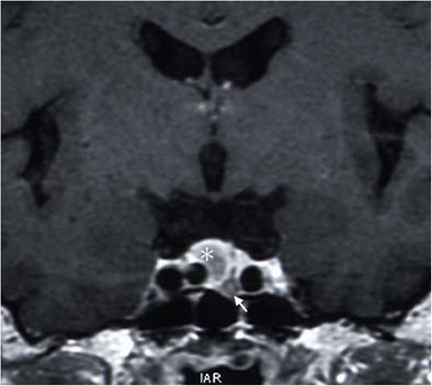
Empty sella see empty sella syndrome.
Double floor of sella turcica.

A Skull X Ray Showed A Double Floor Of Sella B C Magnetic Download Scientific Diagram

Morphological Variants Of Sella Turcica Seen In Lateral Cephalogram A Download Scientific Diagram

Enlarged Sella Turcica Differential Radiology Reference Article Radiopaedia Org

Full Text Association Of Sella Turcica Bridging With Palatal Canine Impaction In Ccide

Figure 1 From Clinical And Radiological Significance Of Sella Turcica A Literature Review Semantic Scholar

Double Contour Of The Floor Download Scientific Diagram
Https Link Springer Com Content Pdf 10 1007 2f978 3 642 67786 1 5 Pdf

Morphologic Variations Of Sella Turcica Classified According To Download Scientific Diagram

Sella Turcica Its Importance In Orthodontics And Craniofacial Morphology Request Pdf

Imaging Of The Cns Ppt Download

Pdf The Size And Morphology Of Sella Turcica A Lateral Cephalometric Study Semantic Scholar
Http Www Neurosurgeryresident Net Onc 20oncology Onc26 20pituitary 20tumors 20apoplexy 20empty 20sella Pdf

Sella And Skull Base Flashcards Quizlet

Pdf Sella Turcica Bridging As A Predicator Of Dentofacial Anomalies A Cephalometric Analysis
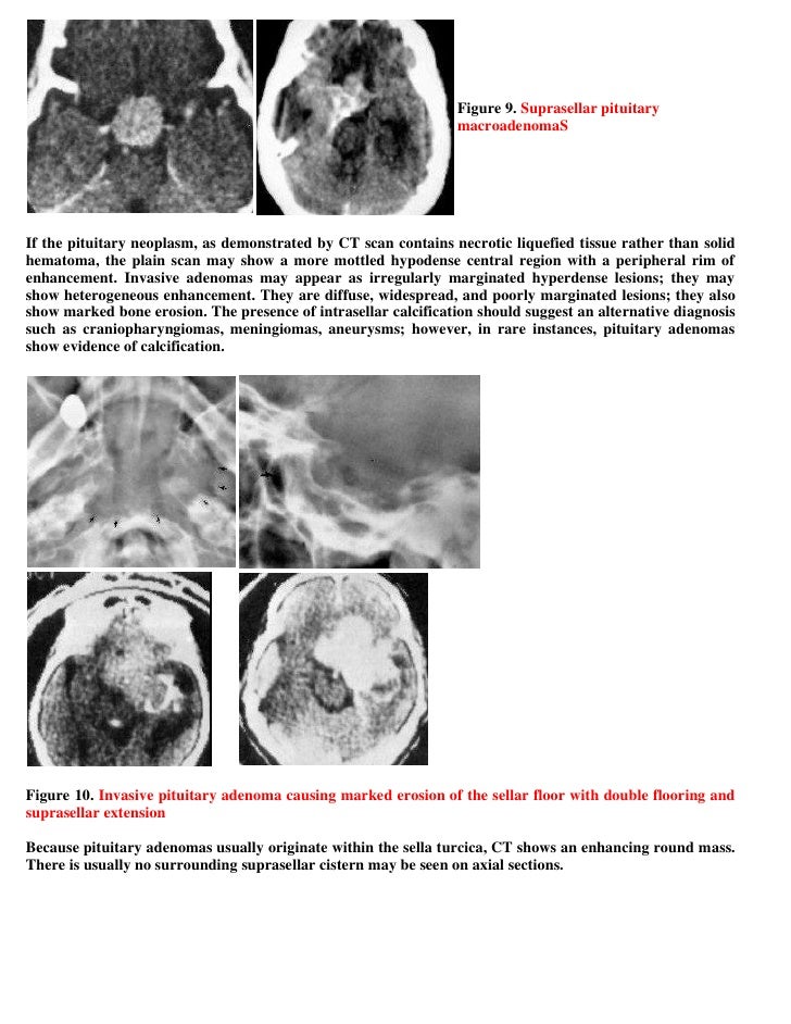
Topic Of The Month Neuroimaging Of Pituitary Adenomas

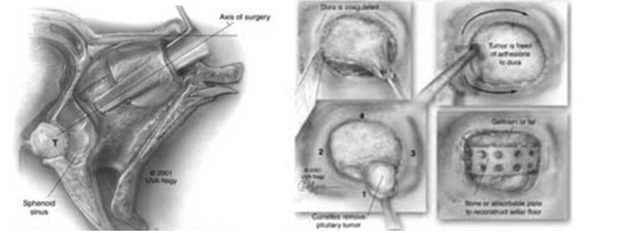
Endoscopic Transplanum And Sellar Approach Sciencedirect
Https Academic Oup Com Neurosurgery Article Pdf 4 Cn Suppl 1 61 20335785 Neurosurgery 4 Cn Suppl 1 61 Pdf

The Use Of Neuroimaging For Assessing Disorders Of Pituitary Development Iorgi 2012 Clinical Endocrinology Wiley Online Library
Https Encrypted Tbn0 Gstatic Com Images Q Tbn 3aand9gcq Eectkp62 Co7iaw99ry63bmp8wmmgpqirwhyw1j0iabak H Usqp Cau
Https Www Birpublications Org Doi Pdfplus 10 1259 0007 1285 48 569 366
Https Journals Viamedica Pl Folia Morphologica Article Download Fm A2019 0042 50362

Sella Turcica An Overview Sciencedirect Topics
Double Pituitary Adenomas Associated With Persistent Trigeminal Artery A Rare Case Report And The Review Of Literature Springerlink

Visual Loss Disorders Of The Chiasm Sciencedirect
Source : pinterest.com
